Simulating complex fMRI-like sources
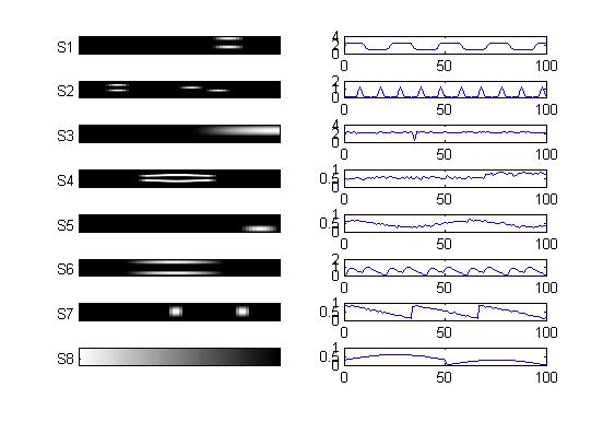
In order to test the performance of separation algorithms for complex-valued data, we generate a simulated complex fMRI-like set of components and mix them with a set of time courses to obtain a simulated fMRI-like dataset. The sources are generated based on the basic knowledge of the statistical characteristics of the underlying neuronal sources. A brief description on the properties of fMRI data is available here.We generate eight complex-valued spatial maps to simulate the fMRI sources and the corresponding time courses. The magnitudes of spatial maps and time courses of each components are as shown in the figure below. In an fMRI experiment, the phase difference induced by the task activation is typically less than pi/9 (see [1, 2]). Therefore, we keep the phase of each pixel uniformly distributed in the range [-pi/18, pi/18]. The phase of each complex-valued time point is generated proportional to its magnitude, but is again restricted to a small range, which in our case is [-pi/18, pi/18]. The spatial sources are arranged into one-dimensional vectors and mixed by the corresponding time courses as the spatial ICA model for fMRI data. References:
- F. G. Hoogenrad, J. R. Reichenbach, E. M. Haacke, S. Lai, K.Kuppusamy, and M. Sprenger, ''In vivo measurement of changes in venousblood-oxygenation with high resolution functional MRI at .95 Tesla by measuring changes in susceptibility and velocity,'' Magn. Reson. Med., vol.39, pp. 97–107, 1998.
- V. D. Calhoun, T. Adali, G. D. Pearlson, P. C. M. van Zijl, and J. J. Pekar, "Independent component analysis of fMRI data in the complex domain," Magnetic Resonance in Medicine, vol. 48, issue 1, pp. 180-192, July 2002.
| Simulated complex fMRI-like sources: |
 |
| Magnitude images of the sources along with their time courses |
Publication in which this set of simulated sources has been utilized:
- W. Xiong, Y.-O. Li, H. Li, T. Adali, and V. D. Calhoun, "On ICA of complex-valued fMRI: Advantages and order selection," in Proc. IEEE Int. Conf. Acoust., Speech, Signal Processing (ICASSP), Las Vegas, Nevada, April 2008. .
Resources:
-
Simulated complex fMRI dataset: Matlab code
